Pyelectasis And Down Syndrome
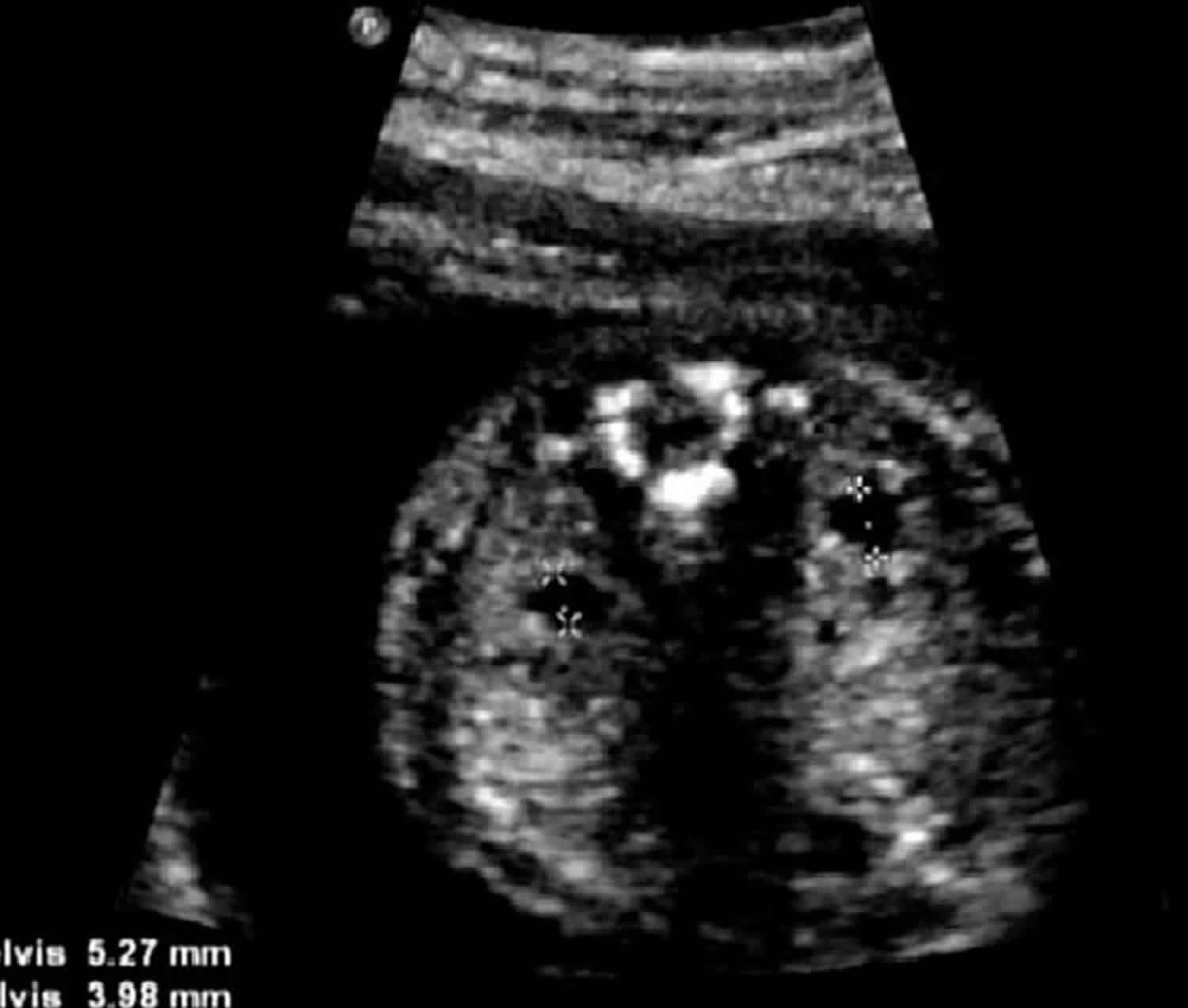
Pyelectasis and down syndrome. Blood tests or amniocentesis are a better way to look for Down syndrome during pregnancy. Benacerraf and colleagues 63 first suggested an association of pyelectasis with aneuploidy primarily Down syndrome in 1990. In a selected high-risk population 25 of.
Pyelectasis was observed in 174 four of 23 of Down syndrome fetuses versus only 2 120 of 5876 of normal controls a statistically significant difference P less than 001. 1 to test the hypothesis that pyelectasis is more common in fetuses with Down syndrome and 2 to determine whether genetic amniocentesis should be offered when dilated renal pelves are identified during fetal ultrasound examination. Ventriculomegaly may be due to other structural abnormalities such as aqueductal stenosis or absence of the corpus callosum or it may be acquired after perinatal infections.
A meta-analysis December 2013. Is the Prevalence Increased for Fetuses With Trisomy 21 September 2006. Coco C Jeanty P.
Ultrasound in Obstetrics and Gynecology Isolated fetal pyelectasis and the risk of Down syndrome. Fetal Pyelectasis or Pelviectasis when seen on ultrasound increases the risk for Down syndrome in the baby by about. Down syndrome can develop in fetus in any pregnancy but the risk increases with maternal age.
However most babies with pyelectasis do not have Down syndrome. Mild renal pyelectasis has an increased risk of aneuploidy particularly trisomy 21 when another risk factor such as advanced maternal age 36 years or associated anomalies are involved 7. The predictive value of pyelectasis for Down syndrome one in 90 compares favorably with other accepted indications for genetic amniocentesis such as advanced maternal age and low maternal serum alpha-fetoprotein.
The most common causes of pyelectasis are. Corteville JE Dicke JM Crane JP. Babies with unresolved pyelectasis may experience urological problems requiring surgery.
No case of trisomy 21 was encountered in the present study but in two cases trisomy 18 was diagnosed. Isolated fetal pyelectasis and chromosomal abnormalities.
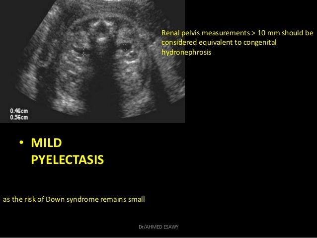
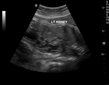
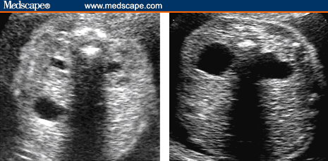
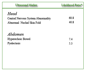
Pyelectasis is considered an ultrasound marker which increases the chance that the baby may have Down syndrome.
Although Down syndrome can occur in any pregnancy the chance for Down syndrome increases with the mothers age. Down syndrome can develop in fetus in any pregnancy but the risk increases with maternal age. Some studies raised concerns about a small risk for Down syndrome with this ultrasound finding. Pyelectasis was observed in 174 four of 23 of Down syndrome fetuses versus only 2 120 of 5876 of normal controls a statistically significant difference P less than 001. However most studies do not find a higher risk for Down syndrome when dilated renal pelvis is the only ultrasound finding. The purpose of this investigation was twofold. Pyelectasis is considered to be a soft marker for Down syndrome. Am J Obstet Gynecol 2005. An abnormal flow of urine from the bladder to the kidneys.
11 rows Isolated fetal pyelectasis is identified in 13 of fetuses during. Am J Obstet Gynecol 2005. Persutte WH Hussey M Chyu J Hobbins JC. Isolated fetal pyelectasis was defined in seven out of 10 studies as a renal pelvis anteroposterior diameter of 4 mm. A blockage of urine between the kidneys and the ureter. Ultrasound in Obstetrics and Gynecology Isolated fetal pyelectasis and the risk of Down syndrome. When pyelectasis is seen on ultrasound the risk for Down syndrome is approximately one and one-half 15 times a womans age-related risk.















Post a Comment for "Pyelectasis And Down Syndrome"